Tumor cells and lung cells both express the pyruvate kinase isoenzyme type M2.
However, the M2-PK isoenzyme found in normal lung tissues is always tetrameric whereas the M2-PK isoenzyme found in tumor cells is mainly dimeric.

The dimeric form of M2-PK is termed Tumor M2-PK.
Differentation between lung M2-PK and Tumor M2-PK
Staining of a moderately to poorly differentiated, non-small cell carcinoma of the left lung lobe
with monoclonal antibodies which specifically recognize the dimeric form of M2-PK (Tumor M2-PK)

Tumor cells show a clear cytoplasmatic Tumor M2-PK staining. Non-malignant cells are not stained with the Tumor M2-PK specific antibody.
Enlargement with intravasal tumor cells

The intravascular Tumor M2-PK staining indicates vascular dissemination of the tumor.
From: Joachim Schneider, Shaker-Verlag Aachen (2003), pp 1-202, ISBN 3-8322-1381-3.
Vmax activities of pyruvate kinase in homogenates of normal lung and lung tumor tissues

Vmax activities were measured spectrophotometrically according to the following
reaction:

Tumor M2-PK concentrations in homogenates of normal lung and lung tumor tissues
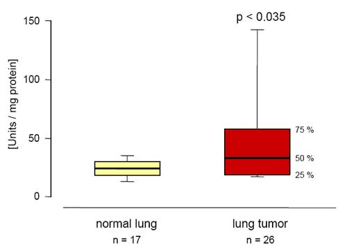
In solid lung tumors pyruvate kinase activities as well as the amount of Tumor M2-PK (dimeric form) increase.
Vmax activities and Tumor M2-PK concentrations in lung control and lung tumor homogenates are part of the doctoral thesis of Anette Beitz, Veterinary Faculty, University of Giessen.
Tumor M2-PK concentrations in EDTA plasma samples from patients with lung tumors Correlation between Tumor M2-PK values and staging
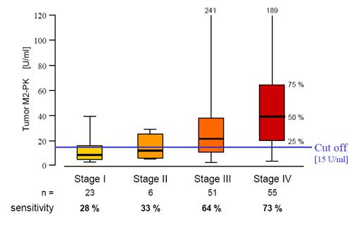
From: J. Schneider (Tumor Markers, AACC Press, Washington, Chapter 47: 471-475).
Tumor M2-PK concentrations in EDTA-plasma samples Follow-up study of a patient with an adenocarcimoma of the lung
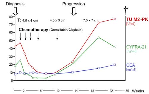
From: Schneider et al.: Anticancer Res. (2000), 20: 5053-5058.
Tumor M2-PK concentrations in EDTA-plasma samples Follow-up study of a patient suffering from small cell carcinoma
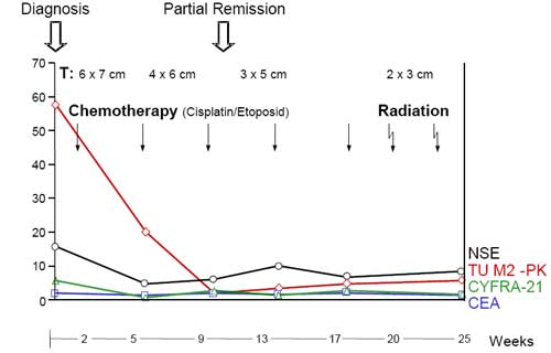
T: radiological tumor size in X-ray
From: Schneider et al.: Anticancer Res. (2002), 22: 311-318.
Tumor M2-PK measurements in EDTA-plasma samples: Joachim Schneider
Tumor M2-PK concentrations were measured with a sandwich ELISA from
ScheBo Biotech AG, Giessen, Germany.